Medispirex
Choosing Mandibular Reconstruction Plates is a clinical decision, not a simple product comparison. The correct plate must support bone continuity, facial symmetry, chewing, speech, and long-term stability. It should also match the defect, graft design, fixation points, and patient-specific anatomy. Small errors can become painful complications.
Professor Rainer Schmelzeisen, a recognized maxillofacial surgeon, has emphasized, “The aim is not only to replace bone, but also to restore function and appearance.” This principle remains highly relevant. A plate may look strong on the operating table, yet fail to provide adequate support after repeated chewing forces. Surgeons therefore assess contour, thickness, screw positioning, material behavior, and compatibility with digital planning. Titanium is widely used because it combines strength, corrosion resistance, and biocompatibility. However, no implant is automatically ideal.
The defect tells the story.
A lateral mandibular defect may require different shaping than a segmental anterior defect. Curved regions near the symphysis demand careful adaptation. Sharp edges, poor seating, or insufficient screw purchase can affect healing and comfort. Patient factors also matter, including radiation exposure, bone quality, infection risk, and planned bone grafting. Yet selection is not always perfectly predictable. Clinical experience can guide the decision, but it cannot replace imaging, surgical judgment, or patient-specific planning. This guide explains how to compare Mandibular Reconstruction Plates thoughtfully, while recognizing their limitations and the need for multidisciplinary review.
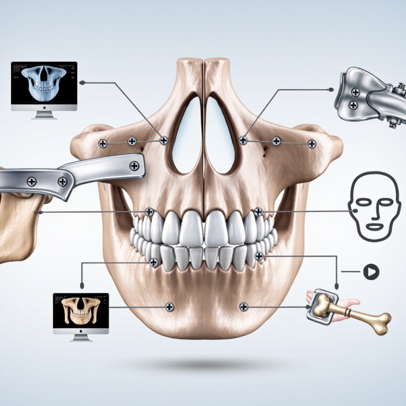
Plate selection should begin with the reconstruction goal, not the plate itself. Ask whether the priority is structural stability, facial contour, oral function, or temporary support. A segmental defect may require stronger load-bearing support than a small continuity defect. Occlusion also matters. Even a well-contoured plate can fail functionally if the mandibular relationship is inaccurate.
Review the patient’s CT images carefully. Note the defect length, remaining bone height, curvature, and available screw positions. Inspect the soft-tissue condition and planned bone graft location. Thin residual bone may limit screw purchase. Dense scar tissue can make adaptation difficult. Previous radiation, infection, or trauma may also change healing expectations. These details should be discussed by the reconstructive surgeon, maxillofacial team, and dental specialists. Their combined assessment is more reliable than a single measurement.
Tips: Map the defect before selecting dimensions. Check plate contour against the planned mandibular shape. Confirm safe screw trajectories near nerves, teeth, and the inferior border. Leave room for soft-tissue coverage. Small errors can become large problems. In practice, goals may change during surgery. I have found that plans based only on digital models can feel too rigid when tissue quality differs from imaging. Reassess the design when clinical findings disagree with the original plan.
Choosing a mandibular reconstruction plate starts with the patient, not the catalog. Review CT images in three dimensions, noting defect length, bone height, curvature, and remaining dentition. Assess the condyle, occlusion, nerve pathways, and soft-tissue coverage before selecting plate geometry. A thin anterior mandible and a broad lateral defect create different mechanical demands. The planned graft and fixation points must match the available bone.
Function should guide the decision. Discuss chewing, speech, swallowing, facial symmetry, and rehabilitation with the reconstructive team. Plate position may affect future dental loading, airway care, or soft-tissue monitoring.
Patients with poor bone quality, previous radiation, active infection, diabetes, or heavy tobacco exposure may face higher complication risks. The team should also examine nutrition, medications, immune status, and follow-up reliability. These details matter clinically.
A rigid construct can protect alignment, but excessive stiffness or prominence may irritate thin soft tissue. Surgeons should compare screw purchase, plate contour, bending needs, and anticipated load changes during healing. Intraoperative findings can challenge the preoperative plan. That is normal. In practice, elegant plans may become unsuitable when bone quality or tissue coverage differs from imaging. Evidence-informed judgment is valuable, but not infallible. Document alternatives, expected risks, and clear triggers for reassessment.
How to Choose Mandibular Reconstruction Plates?
Plate design should follow the defect, not personal preference. Review CT images, occlusion, bone length, and the planned graft before selecting a shape. A load-bearing plate suits large continuity defects or limited bone contact. A lighter bridging design may work when the graft provides reliable support. Consider contouring needs, screw access, soft-tissue coverage, and the risk of plate exposure. Small changes matter.
Material selection also deserves careful judgment. Titanium is widely used because it combines strength, corrosion resistance, and relatively good tissue compatibility. However, strength alone does not guarantee success. A very rigid plate may protect the reconstruction, yet poor contouring can create gaps, pressure points, or unnecessary prominence. I would check the final bend against the mandibular border and avoid repeated bending whenever possible. That step is easy to underestimate.
Fixation should protect both the reconstruction and nearby anatomy. Use preoperative imaging to plan screw length, trajectory, and safe distance from tooth roots, the inferior alveolar canal, and the graft. Locking screws can maintain alignment when bone quality is weak, but they do not replace accurate reduction. Secure fixation near healthy native bone is often more dependable than relying only on fragile graft segments. No strategy is perfect. Intraoperative findings may require a change, especially when soft-tissue coverage or bone purchase is poorer than imaging suggested. A multidisciplinary team should document that decision clearly.
Select the plate design, material, and fixation strategy according to mechanical demands, bone quality, and surgical objectives.
Load-bearing reconstruction plates are preferred when continuity is restored with a segmental graft or when native bone cannot reliably carry occlusal forces. Low-profile or locking designs may help reduce soft-tissue irritation.
Titanium and titanium alloys combine corrosion resistance, biocompatibility, and a lower elastic modulus than stainless steel or cobalt-chromium alloys. The lower modulus may reduce stiffness mismatch with bone.
Use stable bicortical fixation where anatomy permits, distribute screws across healthy bone, and place fixation away from osteotomy lines. Locking screws can preserve plate contour and reduce dependence on plate-to-bone compression.
Representative nominal elastic-modulus values for commonly used implant metals; exact values vary with alloy grade, heat treatment, and manufacturing process. Final selection should follow patient-specific anatomy and validated surgical protocols.
Plate selection should begin with the defect, not a catalogue dimension. Assess the resection length, bone quality, occlusion, soft-tissue coverage, and planned graft. A short plate may fit neatly but leave weak support near the defect. An overly long plate can interfere with flap inset, vessels, or future dental rehabilitation. Computed tomography helps define curvature, available bone, and safe screw trajectories. Clinical judgment remains essential because images cannot show every intraoperative limitation.
Thickness and width should reflect the expected mechanical load. A load-bearing reconstruction may require greater stiffness, especially when bone continuity is absent. A load-sharing situation may permit a slimmer profile when healthy bone contacts are reliable. Hole spacing matters too. It should allow secure fixation on both sides, while avoiding roots, the mandibular canal, and thin cortical regions. Perfect rigidity is not always the best goal. Excessive stiffness can complicate contouring and create prominent hardware. This balance deserves careful review.
Tips: Compare the planned plate with a three-dimensional model before surgery. Check screw length against cortical thickness, not guesswork. Confirm occlusion after provisional fixation. Keep an alternative size available. Measurements can mislead, especially after resection or significant tissue loss. Discuss final dimensions with the reconstructive and maxillofacial teams, using validated mechanical data and local surgical protocols.
Choosing a mandibular reconstruction plate starts with fit, not appearance. A virtual plan should match the patient’s CT anatomy, occlusion, resection line, and planned bone graft. Check the plate along the inferior border, especially near the mandibular angle. A small gap can create a large bending force. AO Surgery Reference recommends stable fixation, adequate screw purchase, and careful soft-tissue coverage. Practical experience also shows that a perfect digital fit may change after resection.
Stability depends on more than screw count. Evaluate bone thickness, screw length, bicortical engagement, plate contour, and load distribution. Reconstruction plates should not obstruct future dental implants or compress delicate soft tissue. Systematic reviews have reported plate exposure rates of roughly 10–20% and plate fracture rates of about 3–8% after vascularized bone reconstruction, although follow-up periods and patient risk factors differ (Journal of Oral and Maxillofacial Surgery, 2021 systematic review). These numbers are warnings, not guarantees.
Compatibility requires checking the fixation system, imaging requirements, instruments, and planned postoperative care. Titanium is widely used, but corrosion concerns, allergy history, radiation exposure, and revision access still deserve review. Long-term assessment should include infection, loosening, fracture, nonunion, and contour changes. I would not accept a low complication rate without examining how the study defined failure. That detail is often overlooked. Patient-specific planning helps, but surgeon judgment remains essential.
| Evaluation Dimension | What to Verify | Practical Assessment | Preferred Characteristics | Potential Concern |
|---|---|---|---|---|
| Anatomical Fit | Whether the plate follows the reconstructed mandibular contour without excessive bending. | Review CT-based planning, patient-specific models, and intraoperative seating along the inferior border and lateral surface. | Close contour conformity, stable seating, and sufficient clearance from the occlusion and soft tissues. | Poor adaptation may introduce gaps, malocclusion, soft-tissue irritation, or unnecessary contouring stress. |
| Reconstruction Geometry | Compatibility with the defect location, number of segments, graft design, and planned mandibular shape. | Confirm plate length, hole distribution, screw access, ramus and symphysis coverage, and the intended osteotomy positions. | Adequate span across the defect with fixation points available in every intended bone segment. | Insufficient span or inaccessible holes can reduce fixation options and compromise alignment. |
| Mechanical Stability | Resistance to bending, torsion, fatigue, and deformation under functional loading. | Compare the plate profile, cross-section, hole design, fixation pattern, and anticipated loading conditions. | High fatigue resistance, appropriate rigidity, and stable fixation without excessive stress concentration. | Overly flexible plates may deform; excessive rigidity may increase stress shielding or concentrate load at plate ends. |
| Screw Fixation | Screw diameter, length, trajectory, locking mechanism, and available bone stock. | Check planned screw paths against CT data, dental roots, the inferior alveolar canal, the graft, and soft-tissue anatomy. | Reliable purchase in viable bone, secure plate-to-screw engagement, and avoidance of critical anatomical structures. | Short purchase length, poor bone quality, or screw collision can reduce construct stability. |
| Material Compatibility | Material composition, corrosion resistance, biocompatibility, and compatibility with the selected screws and graft environment. | Review regulatory documentation, material certificates, implant–screw pairing, and any history of metal sensitivity. | Implant-grade material with documented biocompatibility and a validated matching fixation system. | Unverified material combinations may increase the risk of corrosion, mechanical mismatch, or adverse tissue response. |
| Plate Contouring | Whether the plate can be contoured safely and whether repeated bending is permitted. | Follow the device instructions for bending zones, instruments, bend limits, and intraoperative handling. | Minimal contouring, controlled bending, and no visible damage, sharp edges, or hole distortion. | Repeated or localized bending can create stress risers and reduce fatigue strength. |
| Occlusion and Alignment | Preservation or restoration of mandibular position, facial symmetry, and dental occlusion. | Use preoperative occlusal records, surgical splints when indicated, and intraoperative alignment checks before final tightening. | Stable three-dimensional alignment with reproducible occlusion and appropriate mandibular projection. | Fixing the plate before correcting alignment may lock in malocclusion or asymmetry. |
| Soft-Tissue Compatibility | Plate prominence, edge profile, screw-head position, and relationship to mucosa and skin. | Assess tissue coverage, closure tension, wound vascularity, and areas exposed to mastication or intraoral contamination. | Smooth edges, adequate tissue coverage, and limited prominence in high-risk exposure areas. | Prominence may contribute to wound breakdown, plate exposure, discomfort, or later removal. |
| Imaging and Planning Compatibility | Visibility, artifact profile, and compatibility with CT-based planning and postoperative assessment. | Confirm that the implant can be accurately represented in planning software and assessed on follow-up imaging. | Clear implant identification with manageable imaging artifacts and reliable digital planning data. | Excessive artifact can obscure bone union, screw position, or recurrent disease assessment. |
| Sterilization and Handling | Packaging integrity, sterilization status, shelf life, and handling requirements. | Inspect packaging and implant surfaces, verify the instructions for use, and maintain aseptic handling throughout the procedure. | Intact sterile packaging, traceable documentation, and clearly defined reprocessing or single-use status. | Damaged packaging or unapproved reprocessing may compromise sterility and device performance. |
| Bone and Graft Biology | Relationship between fixation stability, vascularized or non-vascularized grafts, residual bone, and healing potential. | Evaluate bone quality, contact at interfaces, graft vascularity, defect size, radiation history, and infection risk. | A construct that maintains alignment while allowing biological healing and minimizing micromotion. | Poor biology or persistent motion can delay union, cause nonunion, or increase the risk of hardware failure. |
| Long-Term Performance | Evidence of fatigue resistance, union support, complication profile, and need for secondary removal. | Review peer-reviewed clinical evidence, post-market surveillance, follow-up duration, and device-specific adverse-event data. | Documented durability, predictable healing outcomes, and a complication profile appropriate for the patient and defect. | Late loosening, fatigue fracture, infection, exposure, or nonunion may require revision surgery. |
| Documentation and Traceability | Implant identification, lot information, surgical records, and postoperative follow-up requirements. | Record the implant type, dimensions, screw configuration, contouring performed, and postoperative imaging findings. | Complete traceability and clear instructions for future imaging, revision, or removal procedures. | Incomplete records can complicate future treatment, device identification, and complication management. |
Note: Final plate selection, fixation strategy, and surgical technique should be determined by a qualified maxillofacial or reconstructive surgical team according to the patient’s anatomy, diagnosis, bone quality, and applicable device instructions.
The reconstruction goal should guide selection. Stability, facial contour, oral function, or temporary support may have different requirements. Fit comes first.
CT images show defect length, bone height, curvature, and available screw positions. They also reveal thin bone that may limit screw purchase.
A long segmental defect usually needs stronger load-bearing support. A smaller continuity defect may require less structural support. Clinical judgment remains necessary.
Review scarring, tissue thickness, radiation history, infection, and previous trauma. Dense scar tissue can make plate adaptation difficult. Soft-tissue coverage needs space.
A small gap near the inferior border can increase bending forces. Poor contour may affect stability, facial shape, or future dental treatment.
Stability depends on bone thickness, screw length, bicortical engagement, plate contour, and load distribution. More screws do not always mean better fixation.
No. Digital models are useful, but resection findings and tissue quality may differ. Plans can change. Reassess the design when clinical findings disagree.
Follow-up should assess infection, loosening, fracture, nonunion, exposure, and contour changes. Reported complication rates vary between studies and patient groups.
Confirm the fixation system, instruments, imaging needs, and postoperative care plan. Review allergy history, radiation exposure, revision access, and future implant plans. It is not foolproof.
The reconstructive surgeon, maxillofacial team, and dental specialists should review the case together. One measurement rarely captures every clinical risk.
Choosing Mandibular Reconstruction Plates begins with clearly defining the reconstruction goals and evaluating the size, location, and complexity of the mandibular defect. Surgeons should consider the patient’s anatomy, occlusion, soft-tissue condition, functional needs, medical history, and potential clinical risk factors. These assessments help determine the most appropriate plate design, material, and fixation strategy for achieving reliable support and restoring mandibular form and function.
Plate dimensions should correspond to the surgical site, available bone, contour requirements, and expected biomechanical forces. Proper shaping and positioning are essential for stable fixation without interfering with nerves, teeth, joints, or surrounding tissues. Before final placement, the selected Mandibular Reconstruction Plates should be checked for anatomical fit, screw compatibility, structural stability, and resistance to long-term mechanical stress. A careful, patient-specific selection process can improve surgical efficiency, promote predictable healing, and support durable functional and aesthetic outcomes.