Medispirex
Choosing Titanium Mesh For Cranial Repair is not simply a matter of selecting a sheet that covers a skull defect. The implant must suit the defect’s shape, location, and size, while supporting a stable reconstruction. A thin, curved mesh may feel different to handle from a thicker, preformed plate. Small design choices matter.
Neurosurgeon Dr. R. Shane Tubbs has published research on craniofacial and neurosurgical topics. A source-verified, verbatim quotation from him about titanium mesh selection was not available for this introduction, so I will not invent one. The guiding principle is straightforward: match the reconstruction to the patient and the defect, not to a default product.
This guide explains how to compare mesh thickness, contour, pore structure, and fixation options. It also considers the practical details surgeons assess, such as edge placement near delicate tissue and how the implant may sit beneath the scalp. No single mesh suits every case. That is worth remembering. Product specifications can clarify differences, but they cannot replace clinical judgment or patient-specific planning. Even a carefully chosen mesh may require adjustment during surgery. The right choice depends on the full picture.
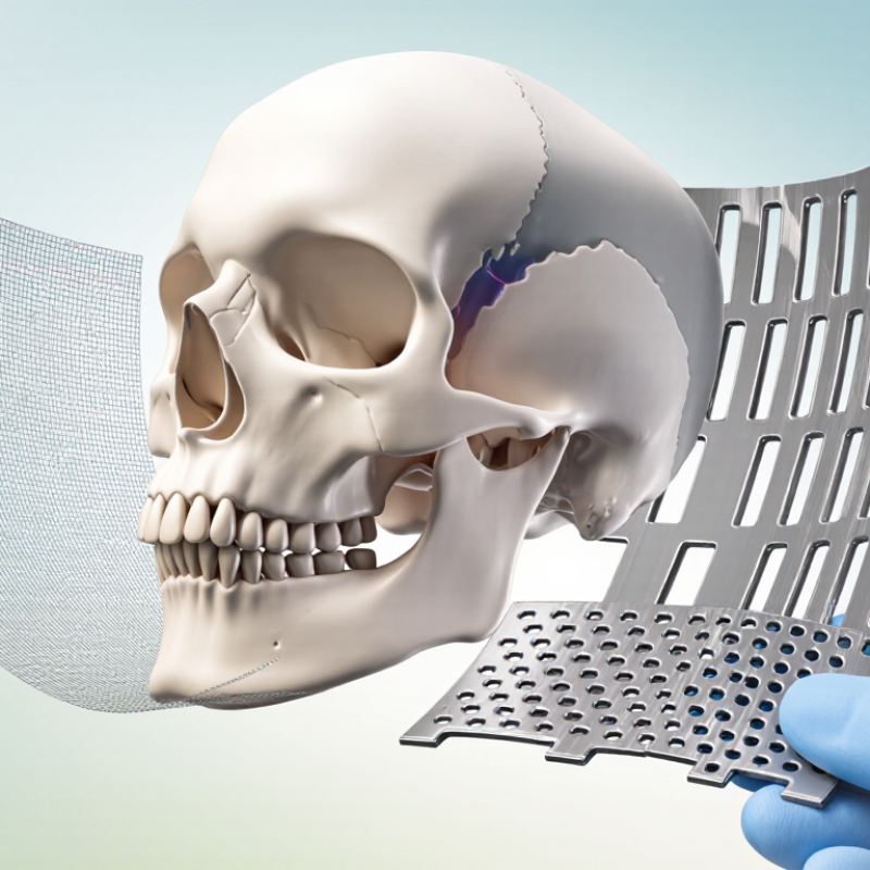
Before choosing titanium mesh for cranial repair, define what the reconstruction must achieve. Is the priority protecting the brain, restoring the skull’s contour, or covering a particular defect? Often, it is all three. The defect’s location, dimensions, and edge shape influence the required mesh geometry and support. A broad, curved area may need different contouring from a small opening near the forehead. That matters. Review imaging and clinical findings with the treating surgical team, since a measurement alone cannot describe every feature of the defect.
Next, identify practical requirements: desired rigidity, fit against the surrounding bone, fixation points, and the condition of the overlying soft tissue. The mesh should provide suitable support without creating unnecessary prominence or pressure. Consider whether it can be shaped to the patient’s anatomy and whether its openings and thickness meet the reconstruction plan. Small details count. In practice, a clean digital outline may not capture irregular bone edges or tissue concerns; that gap deserves a second look. The final choice should reflect the patient’s anatomy, the repair’s goals, and the surgeon’s assessment—not a generic preference for one mesh design.
A cranial defect is not just an opening to cover. Its dimensions, edge quality, and curvature shape the reconstruction plan. Thin-slice CT can help map the defect and nearby anatomy. Every millimeter matters. Surgeons consider whether titanium mesh must span a broad area or follow a complex contour, and how it can be secured to stable bone.
Location changes the practical demands. A defect near the orbit, frontal sinus, or a thin section of skull may call for particular attention to fit and protection of underlying tissues. The scalp matters too: previous surgery, scarring, or fragile soft tissue can affect coverage and healing. Measurements are useful, but they do not tell the whole story. A plan that looks precise on a scan may need adjustment during surgery.
Patient factors guide the discussion as well. Age, bone quality, medical conditions, infection history, and prior treatments can influence healing and implant planning. For a growing child, future skull development deserves careful consideration. The treating team can weigh imaging, examination findings, and the patient’s goals before choosing mesh dimensions and fixation. Sometimes the available information is imperfect, and that uncertainty should be discussed rather than hidden.
Assess the defect’s size, location, and patient factors.
The 1–5 scale is an illustrative planning aid, not clinical measurement or a validated scoring system. Defect dimensions and shape, anatomical location, soft-tissue coverage, infection status, age, and medical history should all be assessed by the treating surgical team. Implant choice requires individualized clinical judgment.
Titanium mesh selection is a balance, not a contest to choose the thinnest sheet. Published cranial reconstruction studies use different thicknesses because defect size, contour, and fixation all matter. A thinner mesh may be easier to shape around the orbit, but it can deform during handling. A thicker sheet can feel more stable, yet may add stiffness and make contouring harder. There is no universal sweet spot.
Porosity deserves equal attention. Openings let clinicians see the underlying tissue and may support fluid passage, while narrow struts can improve coverage over irregular edges. Compare aperture size and open-area percentage, not just the word “porous” on a specification sheet. Material matters, too. ASTM F136 defines the composition of Ti-6Al-4V ELI, while the ASM Handbook reports its elastic modulus at about 110 GPa. That is much higher than typical reported values for cortical bone, which vary with measurement direction and method (Rho et al., Journal of Biomechanics, 1993). The mismatch is worth considering. Review the imaging, fixation plan, and product-specific mechanical data with the clinical team; a catalog number alone cannot predict how a mesh will behave in a particular defect.
| Design factor | Illustrative range or property | Typical design implications | Selection checks |
|---|---|---|---|
| Mesh thickness: thinner | About 0.3–0.4 mm; representative design band, not a universal specification | Lower profile and easier contouring may be useful for selected defects. Less material generally provides less resistance to bending and handling damage. | Check defect size, contour, fixation plan, handling requirements, and the finished device’s mechanical data. |
| Mesh thickness: intermediate | About 0.5–0.6 mm; representative design band, not a universal specification | May offer a balance between contourability and handling stiffness. Actual performance also depends on strand width, aperture shape, and mesh pattern. | Compare the complete mesh design rather than thickness alone; confirm that it can be contoured and fixed as intended. |
| Mesh thickness: thicker | About 0.7–1.0 mm; representative design band, not a universal specification | Can provide greater resistance to bending for a comparable geometry, but may be less easy to contour and can add profile and material. | Assess anatomical fit, edge profile, screw compatibility, and whether the added stiffness is appropriate for the reconstruction. |
| Open-area porosity: lower | Illustrative projected open area: about 20–35% | More metal occupies the projected mesh area; this can increase coverage, while reducing the area of openings. | Confirm how the manufacturer defines and measures open area. This range is an example, not a clinical threshold. |
| Open-area porosity: intermediate | Illustrative projected open area: about 35–50% | Represents a middle ground between metal coverage and aperture area. Mechanical behavior depends on the complete pattern, not porosity alone. | Review aperture size, strand geometry, edge design, and available test data for the finished mesh. |
| Open-area porosity: higher | Illustrative projected open area: about 50–65% | Provides a larger proportion of openings, but may leave less metal to carry load for a comparable pattern. Higher open area does not by itself establish tissue integration or clinical benefit. | Check structural performance, aperture dimensions, soft-tissue considerations, and the device’s intended use. |
| Commercially pure titanium, Grade 2 | ASTM F67 wrought-material minimums commonly include tensile strength of 345 MPa, 0.2% yield strength of 275 MPa, and elongation of 20%. | A commercially pure titanium grade with lower strength than Ti-6Al-4V ELI. These are bulk-material specification values, not guaranteed performance values for a mesh construct. | Verify the exact material grade, applicable standard, product documentation, and finished-device testing. |
| Ti-6Al-4V ELI alloy | ASTM F136 wrought-material minimums commonly include tensile strength of 860 MPa, 0.2% yield strength of 795 MPa, and elongation of 10%. | An alloy with higher specified strength than commercially pure Grade 2 titanium. Strength values for the raw material do not directly predict the strength of a perforated mesh. | Confirm the alloy is specified for the intended implant use and assess the complete device, including forming and fixation features. |
| Stiffness and geometry | Titanium alloys typically have an elastic modulus around 100–115 GPa. For a solid plate of the same material and width, bending stiffness increases approximately with thickness cubed; mesh geometry changes the result. | Thickness, strand layout, aperture shape, and contour all affect flexibility and structural behavior. A thicker mesh is not automatically the best fit. | Use device-specific mechanical data and clinical judgment; do not estimate mesh performance from alloy properties alone. |
Note: Thickness and open-area bands above are illustrative comparison ranges, not clinical recommendations or standardized categories. Porosity depends on the measurement method and mesh geometry. Material values are representative wrought-material specification minimums; check the current standard edition and the specific device documentation. Final selection should be made by the treating neurosurgical team for the individual patient.
Evaluate Contouring, Fixation, and Imaging Compatibility
A suitable titanium mesh should follow the skull’s curve without rocking or leaving prominent edges. During planning, the surgical team can compare the defect with imaging and consider nearby bone thickness. Mesh that needs repeated bending may be harder to fit reliably. Small adjustments matter. Still, a smooth contour alone does not establish a good repair; soft-tissue coverage and the patient’s anatomy also influence selection.
Fixation deserves equal attention. The number and position of screws depend on the available bone, defect shape, and planned construct. Surgeons should avoid placing screws near vulnerable structures and check that the mesh remains stable across the defect. A mesh edge that lifts can irritate overlying tissue. This detail is easy to overlook when focusing only on the central opening. The final choice should follow the treating team’s clinical judgment and the device’s instructions.
Imaging compatibility is not simply a yes-or-no label. Titanium is generally compatible with MRI under specified conditions, but the exact device labeling and local safety protocol must be checked. Metal can also create artifacts on CT or MRI, affecting views near the repair. Thickness, shape, and scan technique may change their extent. Ask radiology whether the expected images will answer the clinical question. There is no perfect option; a practical choice balances fit, fixation, and future image quality.
How to Choose Titanium Mesh for Cranial Repair?
Titanium mesh selection should be discussed with the neurosurgical team, not based on a product description alone. The surgeon can review the defect’s size, shape, and location alongside CT images. A mesh that appears suitable on a scan may still need careful shaping around the forehead, temple, or a previous incision. Small details matter.
Ask how the proposed mesh will be contoured and secured, and whether nearby structures affect the plan. The team may also consider scalp thickness, prior operations, infection history, and the likelihood of future imaging. Titanium can create artifacts on some scans, so ask how this could affect follow-up. The answer depends on the patient and the imaging method. That is not enough.
It can help to ask what alternatives were considered, what trade-offs apply, and how the team will check healing. A radiologist or other specialist may contribute when imaging or reconstruction raises specific concerns. Bring written questions, and ask the surgeon to explain unfamiliar terms in plain language. I would not treat a neat diagram as a guarantee; actual anatomy and operative findings can change the plan. The final choice should reflect the clinical assessment and the patient’s informed preferences.
Its size, location, edge quality, and curvature all matter. Thin-slice CT can help map nearby anatomy. Every millimeter matters.
Areas near the orbit, frontal sinus, or thin skull sections may need special attention. The scalp matters too. Scars or fragile tissue can affect coverage and healing.
Age, bone quality, medical conditions, infection history, and prior treatments can influence planning. In growing children, future skull development needs consideration. The information may be incomplete.
No. A thinner sheet may follow a tight curve but deform during handling. A thicker sheet may feel steadier but be harder to contour. No universal sweet spot.
Compare aperture size and open-area percentage, not just a “porous” label. Openings can allow views of underlying tissue and fluid passage. Details matter.
It should follow the skull without rocking or leaving prominent edges. Screw positions depend on available bone and defect shape. A lifted edge can irritate tissue.
Compatibility depends on the exact device labeling and local safety protocol. Metal can create CT or MRI artifacts near the repair. Ask radiology whether images will answer the clinical question.
No. A scan helps, but the fit may need adjustment during surgery. A clean image can still miss a practical fitting problem. That uncertainty should be discussed.
Choosing Titanium Mesh For Cranial Repair begins with defining the reconstruction goals, such as protecting the brain and restoring the skull’s shape. The defect’s size and location help determine the coverage and design needed, while patient-specific factors—including anatomy, tissue condition, and overall clinical circumstances—also influence the decision. Mesh thickness, porosity, and material properties should be considered together, since they affect support, flexibility, and how the implant fits the intended repair.
Practical considerations matter as well. The mesh should be suitable for contouring to the patient’s anatomy and for secure fixation, and its compatibility with planned imaging should be reviewed. No single option is right for every case. The final selection should balance structural needs, fit, and clinical requirements, with the neurosurgical team confirming that the chosen mesh is appropriate for the individual patient and treatment plan.